Mapping the Brain’s Sense of Gravity
Plute TJ, Spencer DD, Alkawadri R. Age-dependent vestibular cingulate–cerebral network underlying gravitational perception: a cross-sectional multimodal study. Brain Informatics. 2022;9:30. PMID: 36542188.
Gravity is physically constant at the bedside, but our experience of gravity is constructed by the nervous system. In this multimodal study, direct stimulation of anterior and posterior cingulate sites produced a stereotyped sense of floating or weightlessness, creating an unusual window into how the brain integrates vestibular information with perception, action, and the sense of self in space.
The investigation screened 54 intracranial-EEG cases; 10 had cingulate sampling, and 13 stimulation sites entered the Granger-connectivity analysis. Electrical cortical stimulation was combined with passive intracranial connectivity, cortico-cortical evoked potentials, and functional MRI. The converging maps implicated a distributed network linking cingulate regions with the insula, frontal and temporal cortex, parietal regions, brainstem, and cerebellum.
Across those 13 analyzed sites, the paper reported an exploratory association between age at presentation and the degree and extent of Granger connectivity (r = 0.82, multiplicity-corrected p = 0.0035). It is not proof of developmental causation; larger samples are needed before deciding whether higher-order vestibular and cingulate networks follow a longer developmental trajectory than traditional models assume.
The paper also illustrates why no single modality is enough. Intracranial EEG offers exceptional temporal resolution but limited access to subcortical and unsampled structures. fMRI extends the spatial field. CCEP probes effective connectivity. Direct stimulation supplies a causal perturbation. Their overlap is more informative than any one map alone.
Open-access article:
https://doi.org/10.1186/s40708-022-00176-2
PubMed:
https://pubmed.ncbi.nlm.nih.gov/36542188/
Figure caption: Recorded functions, stimulation sites, and Granger-connectivity findings across cingulate regions. Figure 2 from Plute et al., Brain Informatics. 2022;9:30. Licensed under CC BY 4.0; converted to sRGB and resized without changing content.
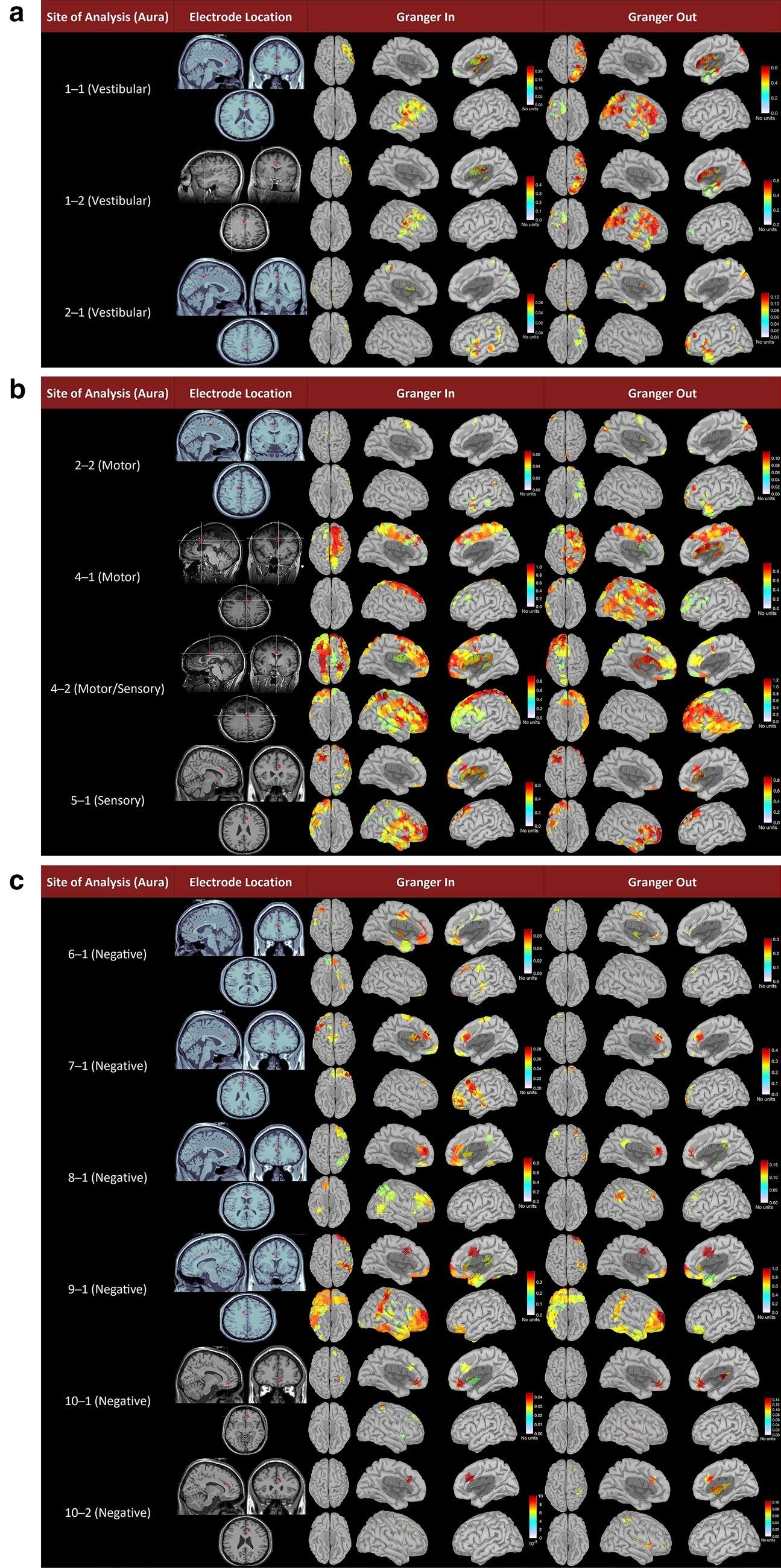
Figure description. Panel A shows three stimulation sites associated with vestibular experiences and their inward and outward Granger-connectivity maps. Panel B shows motor and sensory sites with broader bilateral cortical connectivity. Panel C shows sites with no reported stimulation response, emphasizing that functional effects arise within distributed networks rather than isolated points.
Figure description: Panel A shows three stimulation sites associated with vestibular experiences and their inward and outward Granger-connectivity maps. Panel B shows motor and sensory stimulation sites with broader bilateral cortical connectivity. Panel C shows six sites with no reported stimulation response and their corresponding connectivity maps. Warmer colors indicate stronger connectivity on the figure’s unitless scales.