
Six Minutes of Intracranial EEG: A Proof of Concept for Faster Functional Mapping
Iktimal A, Spencer DD, Alkawadri R. Optimized Multilayer Perceptron for Sensorimotor Functional Mapping Based on a Few Minutes of Intracranial Electroencephalogram Data. Annals of Neurology. 2024;96(1):187–193. PMID: 38506405.
Traditional sensorimotor mapping can require hours of stimulation, observation, and electrophysiological testing. This study asked whether a passive signal already being recorded at the bedside could contribute a useful map much faster.
Using six minutes of free-running intracranial EEG during sleep, the team trained an optimized multilayer perceptron to classify sensorimotor function and identify the anterior lip of the central sulcus. The analysis drew on seven patients and 376 implanted contacts, and reported six performance dimensions—accuracy, area under the curve, recall, precision, F1 score, and specificity—because a clinically useful model must do more than perform well on the majority class.
Feature extension added spectral, connectivity, entropy, and spatial information. Weighting addressed the imbalance between positive and negative contacts. Together, those refinements improved performance compared with base models, particularly on the measures most sensitive to minority-class detection.
The result is a proof of concept for efficient adjunctive mapping, not a replacement for electrical cortical stimulation, somatosensory evoked potentials, imaging, anatomy, or expert review. Its value lies in showing that a few minutes of passive data can contain a recoverable functional signature—and that the way clinical labels and imbalanced data are handled can matter as much as the choice of algorithm.
Full article:
https://doi.org/10.1002/ana.26915
PubMed:
https://pubmed.ncbi.nlm.nih.gov/38506405/
Reproducibility code:
https://github.com/drraf81/CS-Function
Figure caption: Effect of feature extension and class weighting across accuracy, recall, F1 score, and specificity, with a positive relationship between performance changes in central-sulcus and functional classifications. Reused unmodified from the author-accepted-manuscript Figure 5 under Wiley contributor-reuse terms: Iktimal et al., Annals of Neurology. 2024;96(1):187–193.
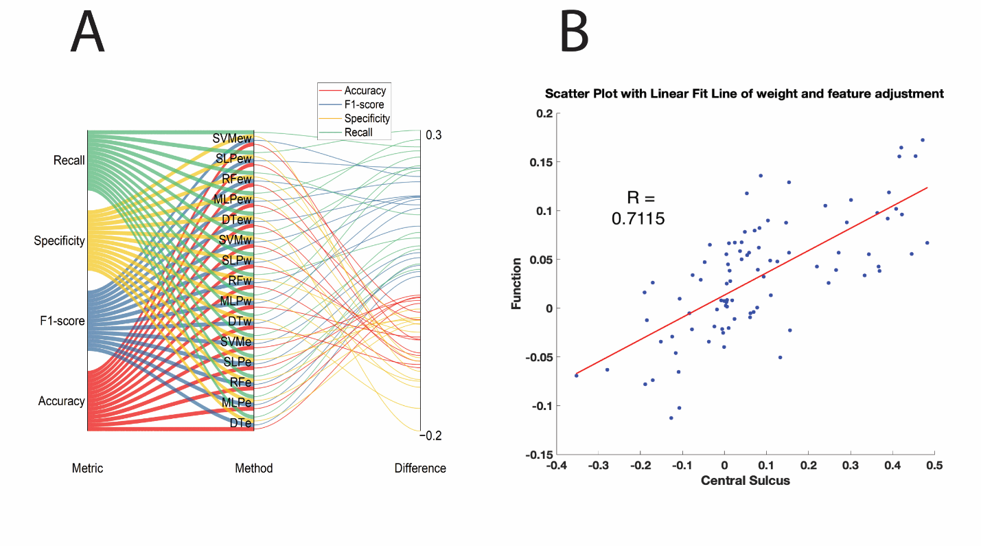
Figure description. Panel A connects four performance metrics to weighted and feature-extended variants of six model families; line width encodes the magnitude of each performance change. Panel B plots optimization gains for central-sulcus classification against gains for functional classification and shows a positive relationship.
Figure description: Panel A connects four performance metrics to weighted and feature-extended variants of six model families. Line width encodes the magnitude of each performance change. Panel B plots mean performance changes for central-sulcus classification against corresponding changes for functional classification; the fitted line slopes upward and the paper reports r = 0.7115 across the models compared in this seven-patient, 376-contact dataset.