Toward a Functional Atlas of the Human Cingulate Gyrus
Mahgoub R, Bayram AK, Spencer DD, Alkawadri R. Functional parcellation of the cingulate gyrus by electrical cortical stimulation: a synthetic literature review and future directions. Journal of Neurology, Neurosurgery & Psychiatry. 2024;95(8):704–721. PMID: 38242679.
The cingulate gyrus is often described as a single limbic structure, but direct electrical stimulation reveals a more intricate landscape. This synthetic literature review assembled 30 studies involving 887 patients and 3,028 implanted contacts to ask a practical question: which human functions can be reproducibly mapped to different cingulate regions? For me, that contrast is the central lesson: a region often treated as functionally quiet or reduced to a single “limbic” label emerges as what I have come to think of as a brain within our brain—the emotional and behavioral brain embedded within the larger brain.
Stimulation produced positive responses at 941 of 3,028 contacts (31.1%). Across the literature, stimulation elicited simple and complex movement, somatosensory experiences, autonomic changes, psychic and affective phenomena, laughter with or without mirth, visual responses, speech effects, and vestibular sensations. The pattern was not a set of hard borders. It was a set of weighted functional tendencies with meaningful overlap.
The anterior cingulate contained much of the affective, laughter, and autonomic response landscape. The middle cingulate carried a strong share of complex motor behavior. The posterior cingulate contributed visual and other higher-order responses while also showing many reproducible null responses. Those nulls matter: an absence of an observed response under a particular stimulation paradigm is evidence about the test as well as the tissue.
The resulting synthesis is clinically useful because electrode implantation and stimulation are both sampling problems. A functional map cannot replace individualized testing, but it can sharpen hypotheses, help align sampling with seizure semiology, and expose where current paradigms are biased toward functions that are easier to elicit or report.
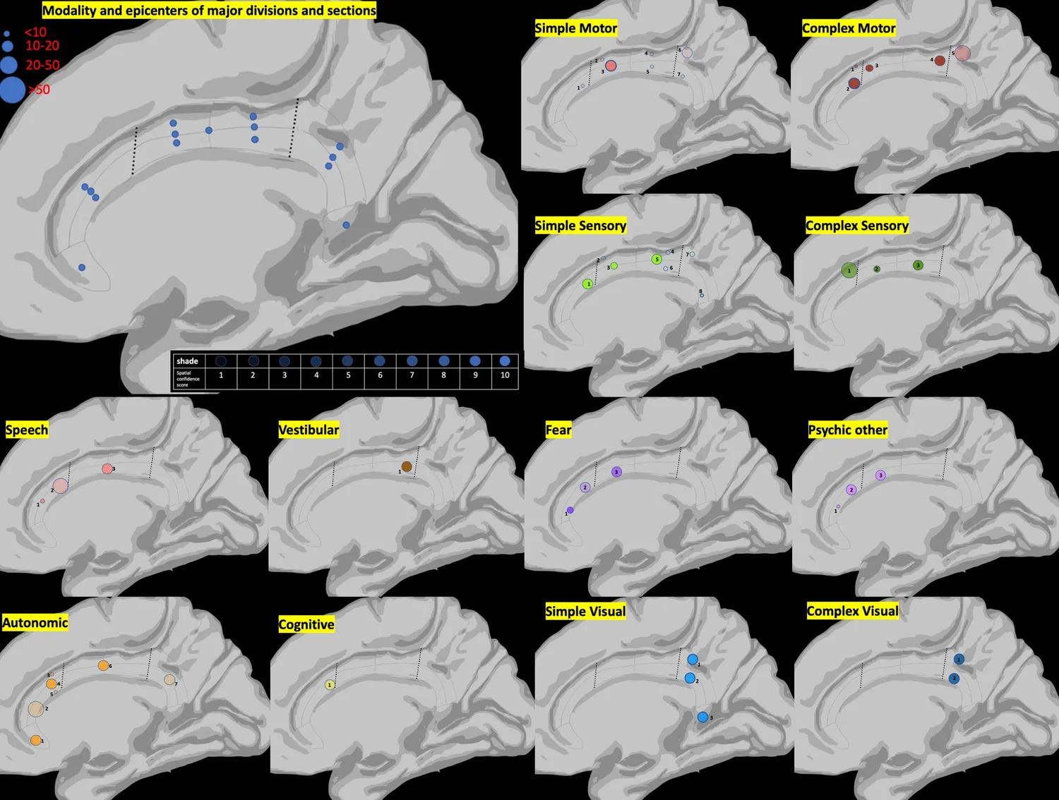
Figure description. The medial brain diagrams summarize reproducible stimulation responses across anterior, middle, and posterior cingulate regions. Larger circles represent more reported cases and darker shading represents greater spatial confidence. Motor and sensory responses concentrate mainly in the middle cingulate, fear and other psychic responses lean anteriorly, and visual responses lean posteriorly, with meaningful overlap rather than hard borders.
Figure description: The medial brain diagrams summarize reproducible stimulation responses across anterior, middle, and posterior cingulate regions. Larger circles represent more reported cases and darker shading represents greater spatial-confidence. Motor and sensory responses concentrate mainly in the middle cingulate, fear and other psychic responses lean anteriorly, and visual responses lean posteriorly, with meaningful overlap rather than hard borders.