The Vision Remains: BCI, AI, and the Slow Work of Changing Epilepsy Care
A perspective piece. The published work discussed here is linked; the assessment of funding, incentives, team science, and organizational structure is my own reading of the field, not a systematic review.
In 2019, our Frontiers in Neuroscience review asked epilepsy care to see intracranial EEG differently: not only as a long recording to be reviewed after the fact, but as a live computational interface. Signals already being collected at millisecond resolution could support real-time seizure detection, functional mapping, network analysis, and decisions that are faster, more reproducible, and more individualized.
In 2022, we revisited that argument on this site for a wider audience. The premise was deliberately practical. A brain–computer interface is not only a futuristic prosthesis. In an epilepsy monitoring unit or operating room, it can be the combination of sensors, continuous data access, computation, and a clinically meaningful output delivered while it can still change a decision.
Seven years after the original review, two truths must be held together. Progress is real, but modest relative to the original vision. Responsive neurostimulation has made closed-loop detection and treatment part of clinical care. Real-time analysis, passive functional mapping, and patient-specific models have continued to advance. Our 2024 Annals of Neurology study showed, as a proof of concept, that six minutes of sleep intracranial EEG can contain enough information to help identify sensorimotor cortex and the anterior lip of the central sulcus.
The larger vision, however, remains incompletely fulfilled. Many epilepsy workflows still depend on prolonged EMU observation, seizure capture, expert visual review, and—when indicated—invasive EEG. Those practices persist for reasons that deserve respect. The stakes are high. Seizures are intermittent, patients are heterogeneous, intracranial sampling is incomplete, and an apparently elegant biomarker can fail when moved from a retrospective dataset to an individual surgical decision. Medicine is slow, sometimes properly so, because its history contains hard lessons about premature certainty.
But caution cannot become a permanent pilot phase. In my experience, translation is slowed by a funding landscape that often rewards novelty in short cycles while underfunding the long work of engineering, multicenter validation, interoperability, maintenance, regulation, and clinical implementation. A prototype can be built by one laboratory. A trustworthy clinical system requires neurologists, neurosurgeons, engineers, data scientists, neuropsychologists, nurses, technologists, ethicists, patients, and product teams to work as one durable unit. It requires collaboration within teams, across institutions, and with industry—not as a late handoff, but from the beginning.
Leadership at the funding level matters just as much. Programs need a longitudinal vision that supports shared protocols, negative and replication studies, protected technical staff, outcome-linked validation, regulatory planning, and the unglamorous infrastructure that keeps a system usable after the grant ends. Funding isolated algorithms without funding the environment in which they must operate produces demonstrations, not transformation.
There is also a subtler organizational layer that is rarely said aloud. People’s livelihoods, professional identities, staffing models, and institutional service structures can become intertwined with long-duration pathways, including EMU admissions and invasive-EEG evaluations. That observation should never be weaponized against the people doing difficult and essential work. It is a responsibility for leadership. A system cannot ask individuals to embrace a shorter or different workflow while making their security depend on the duration and volume of the old one. If technology changes a pathway, the transition must preserve expertise, redesign roles, and return the gains to patients and the teams who care for them.
This is where our 2025 AI-in-neurology editorial meets the earlier BCI vision. AI can help triage large recordings, identify artifacts, expose weak distributed patterns, support passive mapping, integrate modalities, and bring analysis closer to the moment of care. It can also amplify bias, hide failure in rare events behind an accuracy score, and create a new layer of opacity. The standard must therefore be clinical meaning: patient-wise validation, imbalance-aware evaluation, continual monitoring, interpretable failure modes, and accountable clinician–AI partnership.
The goal is not to declare EMU care or invasive EEG obsolete. It is to use every bit of information they produce more intelligently, shorten exposure when evidence truly permits it, reduce repetitive labor, and build less invasive or more preemptive paths where possible. The vision has not failed. It has not yet been fulfilled—and fulfilling it now depends as much on institutions, incentives, and team science as on the next algorithm.
Peer-reviewed 2019 BCI review:
https://doi.org/10.3389/fnins.2019.00191
February 2019 HBM preview:
https://www.humanbrainmapping.net/recent-publications/2019/2/25/brain-computer-interface-bci-applications-in-mapping-of-epileptic-brain-networks-based-on-intracranial-eeg
March 2019 HBM publication note:
https://www.humanbrainmapping.net/recent-publications/2019/3/27/braincomputer-interface-bci-applications-in-mapping-of-epileptic-brain-networks-based-on-intracranial-eeg-an-update
2022 HBM BCI/AI commentary:
https://www.humanbrainmapping.net/blogs/2022/3/10/bci-and-ai-applications-in-epilepsy-care-intracranial-eeg-iceeg-and-neurology
2024 passive-mapping proof of concept:
https://doi.org/10.1002/ana.26915
2025 AI-in-neurology editorial:
https://doi.org/10.3389/fneur.2025.1556510
Figure caption: Hand-motor activation in electrode space using a commonly employed parametric analysis (left) and a custom method designed to improve clinically relevant localization (right); electrical-cortical-stimulation reference contacts are shown in cyan. Figure 3 from Alkawadri, Frontiers in Neuroscience. 2019;13:191. Licensed under CC BY 4.0; converted to sRGB without changing content.
Figure description: Both panels plot the same labeled electrode array. Red circle size represents task-related activation, while cyan labels identify contacts supported by direct electrical cortical stimulation. The left panel contains numerous large activations distributed across the array. The right panel retains a smaller, more selective set near the cyan motor-reference contacts, illustrating why statistical activation and clinically relevant localization are not identical.
Perspective. The published work discussed here is linked; the assessment of funding, incentives, team science, and organizational structure is my own reading of the field, not a systematic review.
From signal stream to live clinical interface
In 2019, our Frontiers in Neuroscience review asked epilepsy care to see intracranial EEG differently: not only as a long recording to be reviewed after the fact, but as a live computational interface. Signals already being collected at millisecond resolution could support real-time seizure detection, functional mapping, network analysis, and decisions that are faster, more reproducible, and more individualized.
In 2022, we revisited that argument on this site for a wider audience. The premise was deliberately practical. A brain–computer interface is not only a futuristic prosthesis. In an epilepsy monitoring unit or operating room, it can be the combination of sensors, continuous data access, computation, and a clinically meaningful output delivered while it can still change a decision.
Seven years after the original review, two truths must be held together. Progress is real, but modest relative to the original vision. Responsive neurostimulation has made closed-loop detection and treatment part of clinical care. Real-time analysis, passive functional mapping, and patient-specific models have continued to advance. Our 2024 Annals of Neurology proof of concept showed that six minutes of sleep intracranial EEG can contain enough information to help identify sensorimotor cortex and the anterior lip of the central sulcus.
Why the vision remains incomplete
The larger vision, however, remains incompletely fulfilled. Many epilepsy workflows still depend on prolonged EMU observation, seizure capture, expert visual review, and—when indicated—invasive EEG. Those practices persist for reasons that deserve respect. The stakes are high. Seizures are intermittent, patients are heterogeneous, intracranial sampling is incomplete, and an apparently elegant biomarker can fail when moved from a retrospective dataset to an individual surgical decision. Medicine is slow, sometimes properly so, because its history contains hard lessons about premature certainty.
But caution cannot become a permanent pilot phase. In my experience, translation is slowed by a funding landscape that often rewards novelty in short cycles while underfunding the long work of engineering, multicenter validation, interoperability, maintenance, regulation, and clinical implementation. A prototype can be built by one laboratory. A trustworthy clinical system requires neurologists, neurosurgeons, engineers, data scientists, neuropsychologists, nurses, technologists, ethicists, patients, and product teams to work as one durable unit. It requires collaboration within teams, across institutions, and with industry—not as a late handoff, but from the beginning.
Leadership at the funding level matters just as much. Programs need a longitudinal vision that supports shared protocols, negative and replication studies, protected technical staff, outcome-linked validation, regulatory planning, and the unglamorous infrastructure that keeps a system usable after the grant ends. Funding isolated algorithms without funding the environment in which they must operate produces demonstrations, not transformation.
There is also a subtler organizational layer that is rarely said aloud. People’s livelihoods, professional identities, staffing models, and institutional service structures can become intertwined with long-duration pathways, including EMU admissions and invasive-EEG evaluations. That observation should never be weaponized against the people doing difficult and essential work. It is a responsibility for leadership. A system cannot ask individuals to embrace a shorter or different workflow while making their security depend on the duration and volume of the old one. If technology changes a pathway, the transition must preserve expertise, redesign roles, and return the gains to patients and the teams who care for them.
Where AI can—and cannot—change the pathway
This is where our 2025 AI-in-neurology editorial meets the earlier BCI vision. AI can help triage large recordings, identify artifacts, expose weak distributed patterns, support passive mapping, integrate modalities, and bring analysis closer to the moment of care. It can also amplify bias, hide failure in rare events behind an accuracy score, and create a new layer of opacity. The standard must therefore be clinical meaning: patient-wise validation, imbalance-aware evaluation, continual monitoring, interpretable failure modes, and accountable clinician–AI partnership.
The goal is not to declare EMU care or invasive EEG obsolete. It is to use every bit of information they produce more intelligently, shorten exposure when evidence truly permits it, reduce repetitive labor, and build less invasive or more preemptive paths where possible. The vision has not failed. It has not yet been fulfilled—and fulfilling it now depends as much on institutions, incentives, and team science as on the next algorithm.
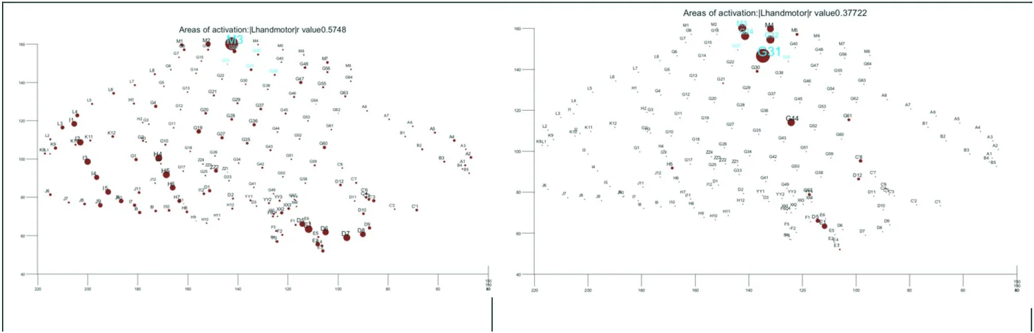
Figure description. Both panels plot the same labeled electrode array. Red-circle size represents task-related activation, while cyan labels identify contacts supported by direct electrical cortical stimulation. The conventional analysis shows broad activation; the custom analysis retains a smaller set concentrated near the motor-reference contacts.